

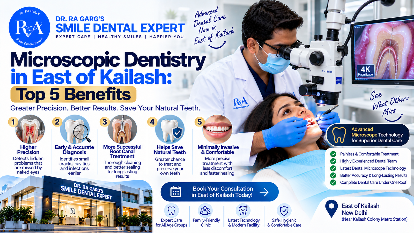
Microscopic dentistry in East of Kailash helps dentists examine teeth and root canals under magnification for more detailed diagnosis and treatment planning. At Smile Dental Care Centre, patients can learn whether microscope-assisted dentistry is suitable for their dental condition.
What are the top 5 benefits of microscopic dentistry? In one line: it lets your dentist see at up to 20–25x magnification what the naked eye simply cannot — hidden canals, hairline cracks, and leftover decay — which translates into more accurate diagnoses, more conserved natural tooth, and treatments that last years longer. Microscopic dentistry, especially microscopic root canal treatment, is quickly becoming the benchmark for quality dental care in East of Kailash and South Delhi. At our Captain Gaur Marg, Kailash Hills clinic, patients from Greater Kailash, Kailash Colony, Lajpat Nagar, Nehru Place and C.R. Park — and from Faridabad’s Sector 34 at our second branch — increasingly ask us about it by name. This guide explains what the dental operating microscope actually does, the five biggest benefits backed by clinical logic, and when magnification makes a genuine difference to your treatment outcome.
Table of Contents
- What Is Microscopic Dentistry?
- Benefit 1: Diagnosis That Catches What X-Rays and Eyes Miss
- Benefit 2: No Missed Canals in Microscopic Root Canal Treatment
- Benefit 3: Maximum Preservation of Natural Tooth
- Benefit 4: Fewer Failures, Fewer Re-Treatments
- Benefit 5: Documentation You Can Actually See
- Which Procedures Benefit Most from the Microscope?
- Microscope vs Naked-Eye Dentistry: Quick Comparison
- Microscopic Dentistry Cost in East of Kailash & South Delhi
- FAQs
- Our Clinic Locations
What Is Microscopic Dentistry?
Microscopic dentistry means performing dental procedures under a dental operating microscope (DOM) — a ceiling- or floor-mounted instrument that magnifies the working field 3x to 25x and floods it with shadow-free LED illumination. To picture the difference: the pulp canal openings inside a molar can be smaller than 0.5 mm, roughly the tip of a ballpoint pen. At normal vision, a dentist works on them partly by feel and experience. Under 20x magnification, each opening is as visible as a coin on a table.
Microscopes transformed fields like neurosurgery and ophthalmology decades ago, and endodontics (root canal science) adopted them for the same reason: outcomes improve when the surgeon can actually see the detail they are treating. Research catalogued by the National Institute of Dental and Craniofacial Research (NIDCR) consistently links enhanced visualisation and magnification to more complete canal debridement — the single biggest factor in root canal success.
Microscope vs loupes: not the same thing
Many clinics advertise “magnification” while using loupes — spectacle-mounted lenses of 2.5x–4x. Loupes are useful, but a dental operating microscope offers 5–6 times more magnification, coaxial lighting that looks straight down the canal, and stable, fatigue-free ergonomics for long procedures. When a clinic says microscopic dentistry, ask to see the microscope.
Benefit 1: Diagnosis That Catches What X-Rays and Eyes Miss
Many of dentistry’s most frustrating problems are nearly invisible. Cracked tooth syndrome — unexplained pain on chewing — is a classic example: the fracture line is often too fine for X-rays and too subtle for the naked eye. Under the microscope, hairline cracks, micro-leakage around old fillings, early decay at the margins of crowns, and perforations show up clearly.
This changes real decisions. A tooth with a crack limited to the crown portion can be saved with a protective cap; a crack running down the root means extraction and replacement planning. Guessing wrong in either direction costs the patient either a tooth or thousands of rupees — magnified diagnosis takes the guesswork out. It also prevents over-treatment: a stained groove that looks like decay to the eye is often confirmed as sound enamel at 16x, meaning no drilling at all.
Benefit 2: No Missed Canals in Microscopic Root Canal Treatment
The number one cause of root canal failure is anatomy the dentist never saw. Upper first molars hide a second canal (the famous MB2) in more than half of cases; lower incisors often carry two canals instead of one; some premolars split into three. A canal that is never found is never cleaned — and the bacteria left inside quietly rebuild the infection over months or years.
Under the operating microscope, the pulp floor’s subtle colour map — dark developmental lines pointing to hidden openings — becomes readable. Calcified canals that have shrunk with age can be located and negotiated without gouging the tooth. This is the core promise of microscopic root canal treatment: every canal found, cleaned to full length, and sealed. It is a natural upgrade to the comfort-focused approach we already follow in our painless root canal treatment for Delhi and Faridabad patients, and the standard we apply in complex cases for our root canal treatment in East of Kailash practice — convenient for patients from Greater Kailash, Lajpat Nagar and C.R. Park.
Benefit 3: Maximum Preservation of Natural Tooth
Modern dentistry’s guiding principle is minimal intervention: remove only what is diseased, keep everything that is healthy. The American Dental Association’s MouthHealthy guidance repeatedly emphasises that natural tooth structure, once removed, never grows back.
Working at high magnification lets the dentist honour that principle literally millimetre by millimetre. Access openings for root canals can be kept conservative because the operator can see inside through a smaller window. Decay removal stops precisely at the junction of soft, infected dentine and hard, healthy tissue instead of taking a wide safety margin. Old filling replacement removes the filling, not the surrounding tooth. The long-term payoff is substantial: teeth with more remaining natural structure resist fracture better, need crowns less often, and survive decades longer.
Benefit 4: Fewer Failures, Fewer Re-Treatments
Every re-treatment is a cost the first treatment failed to prevent — in money, time, and tooth structure. Microscopic technique attacks the three most common failure sources at once: missed anatomy (found and treated), incomplete cleaning (verified visually at high magnification before sealing), and poor sealing (margins of fillings and crowns checked at magnification rather than by feel).
For patients this shows up as fewer surprises at the one-year review X-ray, fewer “the pain came back” phone calls, and root-canal-treated teeth that comfortably carry their crowns for 10–20 years. It also expands what is saveable: separated instrument fragments can be retrieved, perforations repaired, and calcified canals negotiated — teeth that would once have been extracted and replaced can often be rescued. Considering a single dental implant in Delhi costs ₹25,000–₹50,000 in 2026, every tooth the microscope saves is a significant financial win too.
Benefit 5: Documentation You Can Actually See
Dental operating microscopes carry integrated HD cameras, which means your treatment can be photographed and recorded through the exact lens the dentist works through. For patients this creates a new level of transparency: you can see the crack we are describing, the cleaned canals before sealing, and the final result — not as an abstract X-ray, but as a real image of your own tooth.
This matters practically. Before-and-after records protect you when seeking insurance reimbursement, give any future dentist a precise history, and let you verify you received what you paid for. We consider visible proof part of honest dentistry, alongside the written estimates and X-ray sharing we practise at both branches — see what else we have invested in on our facilities page.
Which Procedures Benefit Most from the Microscope?
- Root canal treatment — locating all canals, navigating curves and calcifications, verifying complete cleaning.
- Root canal re-treatment — removing old filling material and finding what the first attempt missed.
- Cracked tooth diagnosis — tracing fracture lines to decide if a tooth is saveable.
- Precision fillings and crown margins — sealing restorations so bacteria cannot leak underneath; this complements the aesthetics of tooth-colored fillings.
- Apical microsurgery — treating stubborn root-tip infections surgically with tiny, precise access.
- Instrument retrieval and perforation repair — rescue procedures that are nearly impossible without magnification.
Microscope vs Naked-Eye Dentistry: Quick Comparison
| Parameter | Microscopic Dentistry | Naked Eye / Loupes |
|---|---|---|
| Magnification | 3x – 25x with coaxial light | 1x (eye) / 2.5x–4x (loupes) |
| Hidden canal detection (e.g. MB2) | High — visualised directly | Depends on feel and experience |
| Crack diagnosis | Fracture lines traced visually | Often missed or found late |
| Tooth structure removed | Minimal, precision-guided | Wider safety margins needed |
| Verification of cleaning | Visual confirmation before sealing | Indirect (X-ray and tactile only) |
| Procedure records | HD photos/video through the lens | X-rays only |
| Typical RCT success benefit | Higher, especially molars/re-treatments | Good for simple anatomy |
Microscopic Dentistry Cost in East of Kailash & South Delhi
Does it cost more? Modestly, yes. A dental operating microscope is a multi-lakh investment, and microscope-assisted procedures take more chair time because the work is more thorough. At our East of Kailash clinic and across South Delhi in 2026, expect microscope-assisted root canal treatment to run roughly ₹1,500–₹4,000 above standard pricing — typically ₹6,500–₹12,000 for a molar, against ₹5,000–₹8,000 conventionally. Re-treatments and microsurgery range ₹8,000–₹15,000.
Weigh that premium against what it prevents: a failed RCT means paying again for re-treatment (₹6,000–₹10,000), a new crown, or — worst case — extraction plus an implant. For complex molars, calcified canals, and previously failed treatments, magnification is not a luxury; it is the difference between hoping and knowing. For simple front teeth with straight single canals, conventional treatment by an experienced hand remains perfectly sound — and we will tell you honestly which category your tooth is in. Explore everything our dental clinic in South Delhi offers under one roof.
Frequently Asked Questions
What is microscopic root canal treatment?
It is root canal therapy performed under a dental operating microscope at up to 20–25x magnification. The dentist can see every canal opening, clean curved and hidden canals completely, and visually verify the result before sealing — significantly improving accuracy over naked-eye treatment.
Is microscopic dentistry painful?
No — if anything it is more comfortable. The procedure still uses standard local anaesthesia, but greater precision means less drilling, smaller access openings, and less post-operative soreness than conventional technique.
How much does a microscopic root canal cost in East of Kailash, South Delhi?
Roughly ₹6,500–₹12,000 for a molar in 2026 at our East of Kailash clinic, about ₹1,500–₹4,000 more than a conventional RCT. Re-treatments under the microscope range ₹8,000–₹15,000, with identical pricing at our Faridabad branch. You receive a written estimate before treatment begins.
Is the microscope necessary for every root canal?
Not every one. Simple front teeth with single straight canals do well with conventional technique. The microscope earns its premium in molars, calcified canals, suspected cracks, and failed previous root canals — cases where unseen anatomy decides success.
Can a failed root canal be saved with microscopic re-treatment?
Very often, yes. The microscope lets the dentist find the missed canal or leak that caused the failure, clean it properly, and reseal. Many teeth scheduled for extraction elsewhere are rescued this way — always worth a second opinion before removing a tooth.
Which doctor handles microscopic and surgical cases at Smile Dental Care Centre?
Complex endodontic and surgical cases are overseen by Dr. Ashish Garg, our oral and maxillofacial surgeon, supported by our endodontic team and digital imaging at the East of Kailash (Kailash Hills) clinic — a short drive from Greater Kailash, Nehru Place and Kailash Colony — and at our Faridabad branch.
Our Clinic Locations
Smile Dental Care Centre — East of Kailash, New Delhi
C 8, Captain Gaur Marg, C Block Kailash Hills, East of Kailash, New Delhi 110065
Phone: 011 35966954 / +91 9318339020
Get directions on Google Maps (East of Kailash)
Smile Dental Care Centre — Ashoka Enclave, Faridabad
210, G Floor, Ashoka Enclave Main, Sector 34, Opp. Devsthal Mandir, Near Sarai Metro Station, Faridabad 121001
Phone: 0129 412 8720 / +91 99990 27270
Get directions on Google Maps (Faridabad)
Want to know if your case needs the microscope? Send your X-ray on WhatsApp +91 93183 39020 for an honest assessment.
See the Difference Precision Makes
If you have a complicated molar, a root canal that failed elsewhere, or unexplained pain on chewing, precision diagnosis is the smartest first step. Call +91 93183 39020, reach us on WhatsApp, or book a consultation online at Smile Dental Care Centre, East of Kailash or Faridabad — and let’s save the tooth you were told couldn’t be saved.
